SnapGene Design of a Frataxin-AcGFP1 Fusion Vector with XhoI and PstI Cloning
Published:
Design a vector expressing human frataxin with GFP fused to its C-terminus, plus an experiment to find where frataxin goes inside human cells.
Primer design
Working from the frataxin coding sequence (isoform 1 preproprotein, cDNA IMAGE:4842134 in pOTB7), with EMBOSS Sixpack used to resolve the reading frames:
- Forward
ATGTGGACTCTCGGGCGCCGCGCAG, Tm 67.5 C - Reverse
AGCATCTTTTCCGGAATAGGCCAAGGAAGAC, Tm 63 C - The stop codon is deliberately omitted so that translation reads on into GFP
- Predicted product 630 bp, amplified at 95 C for 2 min, then 30 cycles of 95 C 20 s, 58 C 20 s and 72 C 15 s, with a 3 min final extension
Choosing the enzymes
Thirteen enzymes in the pAcGFP1-N1 multiple cloning site were screened against the frataxin sequence itself. BglII, SacI, SacII and SmaI all cut inside the gene and were ruled out, which is the check that decides the whole design: an enzyme that cuts the insert destroys it.
Of five workable pairs, XhoI and PstI were chosen, both active in Buffer H so the digest can be done in one reaction. Each primer carries a GGG clamp plus its site (CTCGAG or CTGCAG) ahead of the gene-specific sequence, the clamp being there so the enzyme has enough flanking DNA to bind and cut.
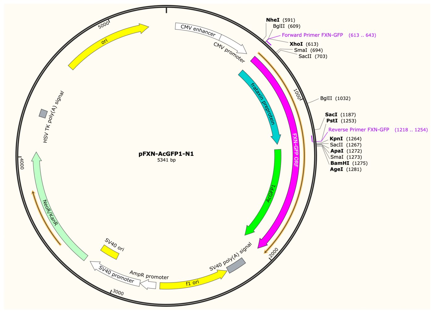
The designed vector pFXN-AcGFP1-N1 (5,341 bp) in SnapGene: frataxin fused to AcGFP1 under the CMV promoter, cloned between XhoI and PstI, with both primers marked.
Checking the frame
A C-terminal fusion only works if frataxin’s codons run into GFP’s without a stop and without a frameshift. One base out at either junction and the construct expresses frataxin followed by nonsense, or nothing fluorescent at all. Both junctions were therefore checked explicitly in SnapGene.
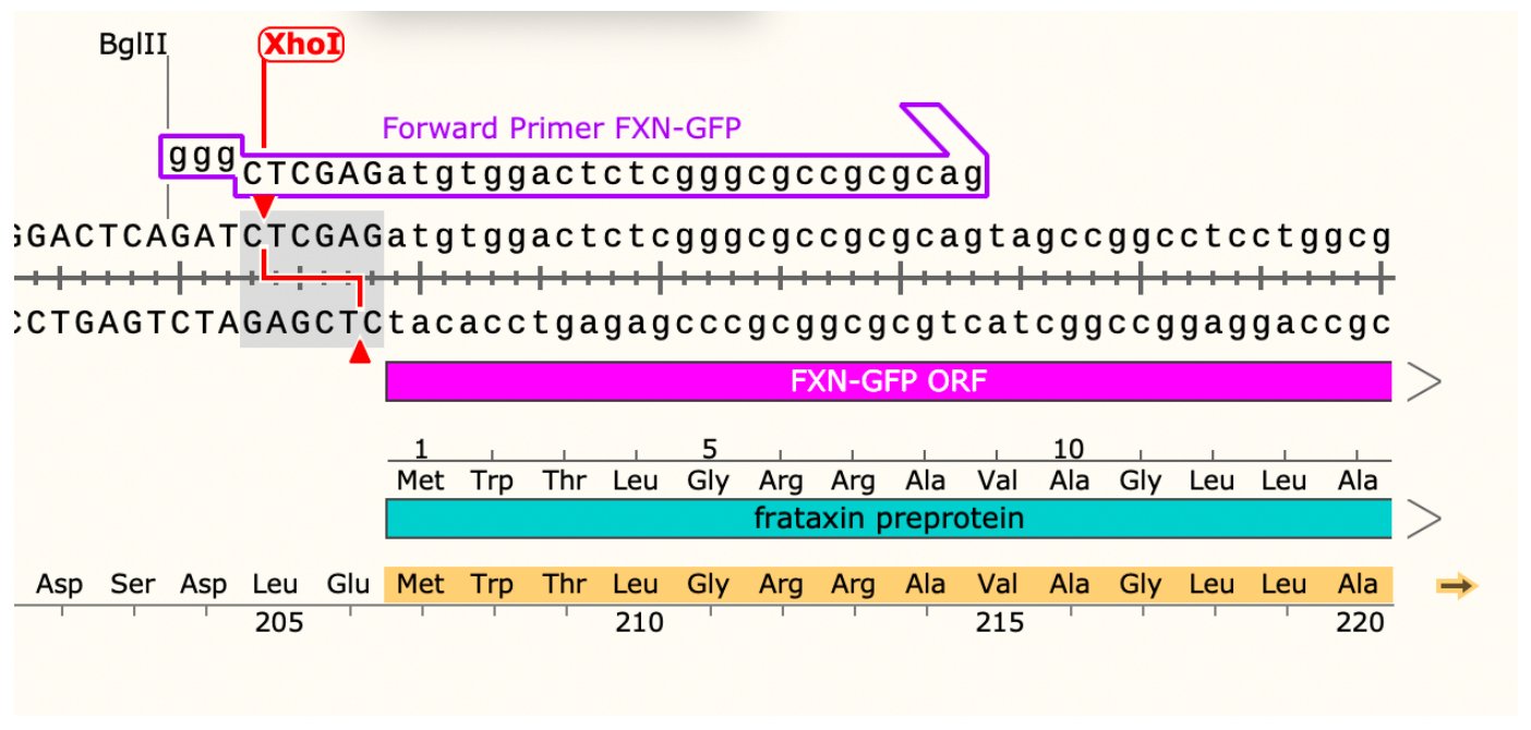
Start of the fusion: the forward primer’s GGG clamp and XhoI site at the frataxin start codon.
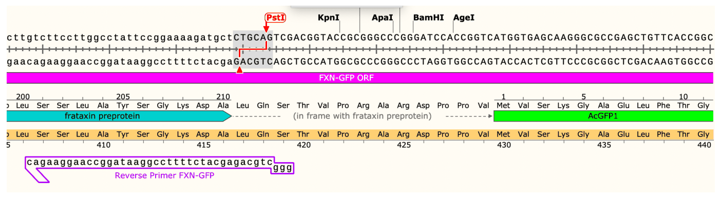
The frataxin to AcGFP1 junction: the PstI site and reverse primer, with GFP in frame.
The cloning plan and the experiment
The full route was written as a flow diagram: PCR, digestion, dephosphorylation, ligation, transformation, colony screening, and confirmation by NheI and BamHI digestion followed by sequencing.
The biological experiment: electroporate the plasmid into HeLa cells, select with G418, then image frataxin-GFP by fluorescence microscopy at roughly 475 nm excitation and 505 nm emission. Frataxin is expected to localise to mitochondria.
These plasmid maps are designs made in SnapGene rather than constructs that were built and verified.