
AMBER99 Energy Minimisation and Manual Docking of Antifolates into DHFR
Published:
Molecular mechanics and manual docking in YASARA (Krieger, Koraimann & Vriend, Proteins 47, 393-402), using the AMBER99 force field and steepest-descent minimisation.
Conformational energies
The first half establishes that structure has an energetic cost, by measuring it:
- Why left-handed helices are rare. A right-handed alpha helix of L-alanines scored 1,520.54 kJ/mol against 1,979.93 kJ/mol for the D-alanine equivalent.
- Why the Ramachandran plot has forbidden regions. An alanine dipeptide at a disallowed phi/psi point cost over 20,000 kJ/mol, against roughly 15 to 130 kJ/mol at the three allowed points. The blank areas of a Ramachandran plot are steric impossibility, not convention.
- Ethane, eclipsed at 20.15 kJ/mol against staggered at 7.56 kJ/mol after minimisation.
These are single force-field values from one program, useful for comparing conformations against each other rather than as absolute energies.
Dihydrofolate reductase and methotrexate
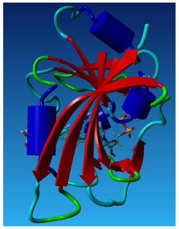
Dihydrofolate reductase bound to methotrexate (PDB 3DFR), the eight-stranded beta sheet in red and ligands as sticks.
Mapping the interactions gave hydrogen bonds from the pteridine ring to Asp26, Thr116, Leu4 and Ala97 plus two through bridging waters, an electrostatic contact with Asp26, stacking against a tryptophan, and the glutamate tail held by His28 and Arg57.
Trimethoprim: bound conformations are strained
Minimising trimethoprim from a high-energy start reached -1,354.90 kJ/mol, a local rather than global minimum on the published energy surface. The crystal conformation scored -1,055.98 kJ/mol and the DHFR-bound conformation -990.36 kJ/mol. The drug binds in a shape it would not adopt on its own, and the binding contacts are what pay for the strain.
Docking by hand
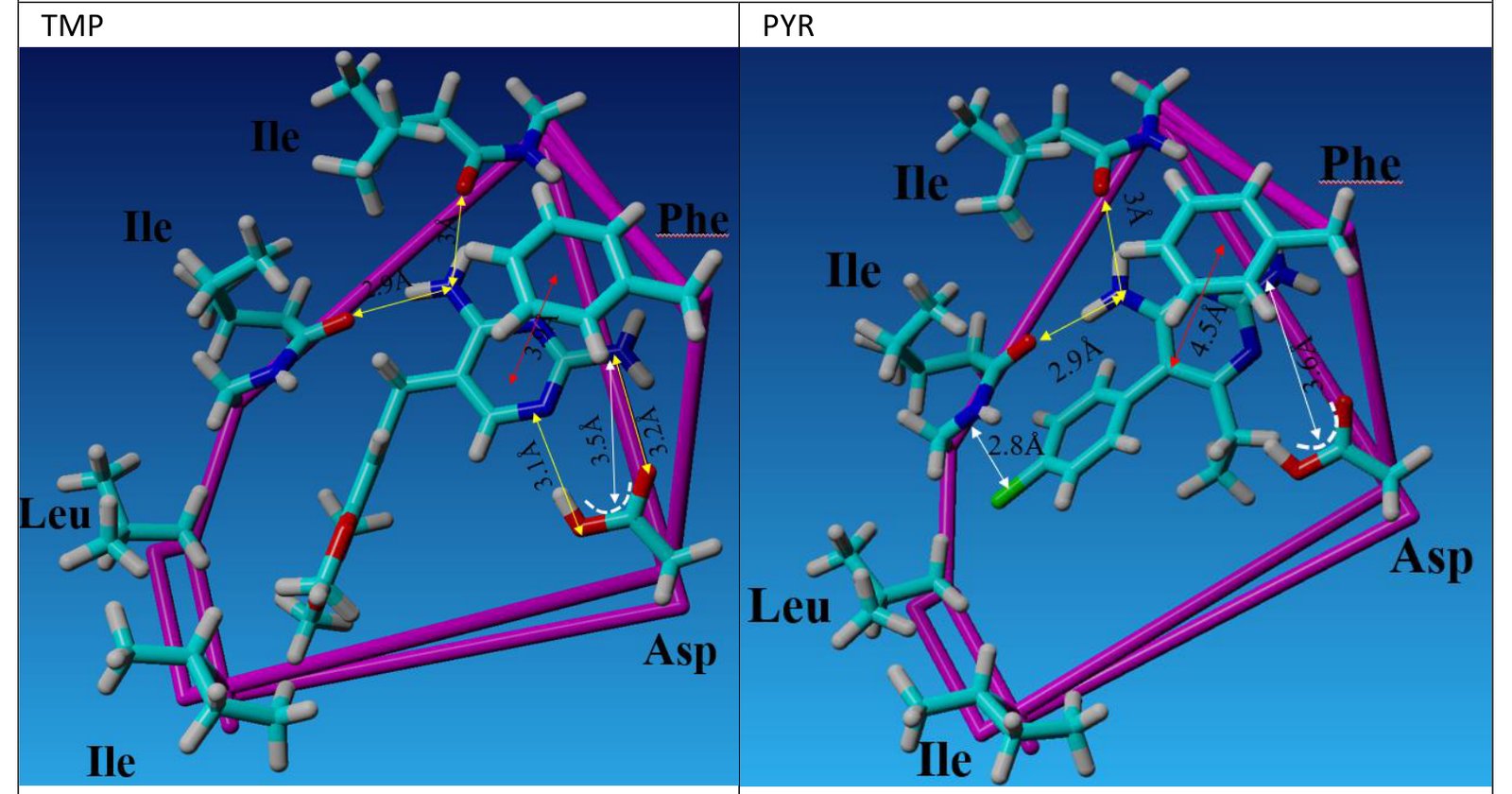
Trimethoprim (left) and pyrimethamine (right) manually docked into a simplified active site in YASARA. Arrows mark hydrogen bonds (yellow), electrostatic contacts (white) and ring stacking (red), with distances in angstroms.
Trimethoprim docked into a simplified site built from an aspartate, a phenylalanine, a leucine and three isoleucines made four hydrogen bonds of 2.8 to 3.2 Angstroms, two of them to the aspartate, plus an electrostatic contact at 3.5 Angstroms and stacking with the phenylalanine at about 4 Angstroms. That dependence on the aspartate is consistent with the reported 30-fold loss of affinity when it is mutated to asparagine.
Pyrimethamine, built and minimised separately (-1,413.90 kJ/mol), cannot adopt the same pose. It has less rotational freedom and its ethyl group clashes with the aspartate, so it loses both hydrogen bonds to it (3.6 Angstroms), while its chlorine adds contacts elsewhere. The overall affinity comes out similar to, or slightly above, trimethoprim’s.
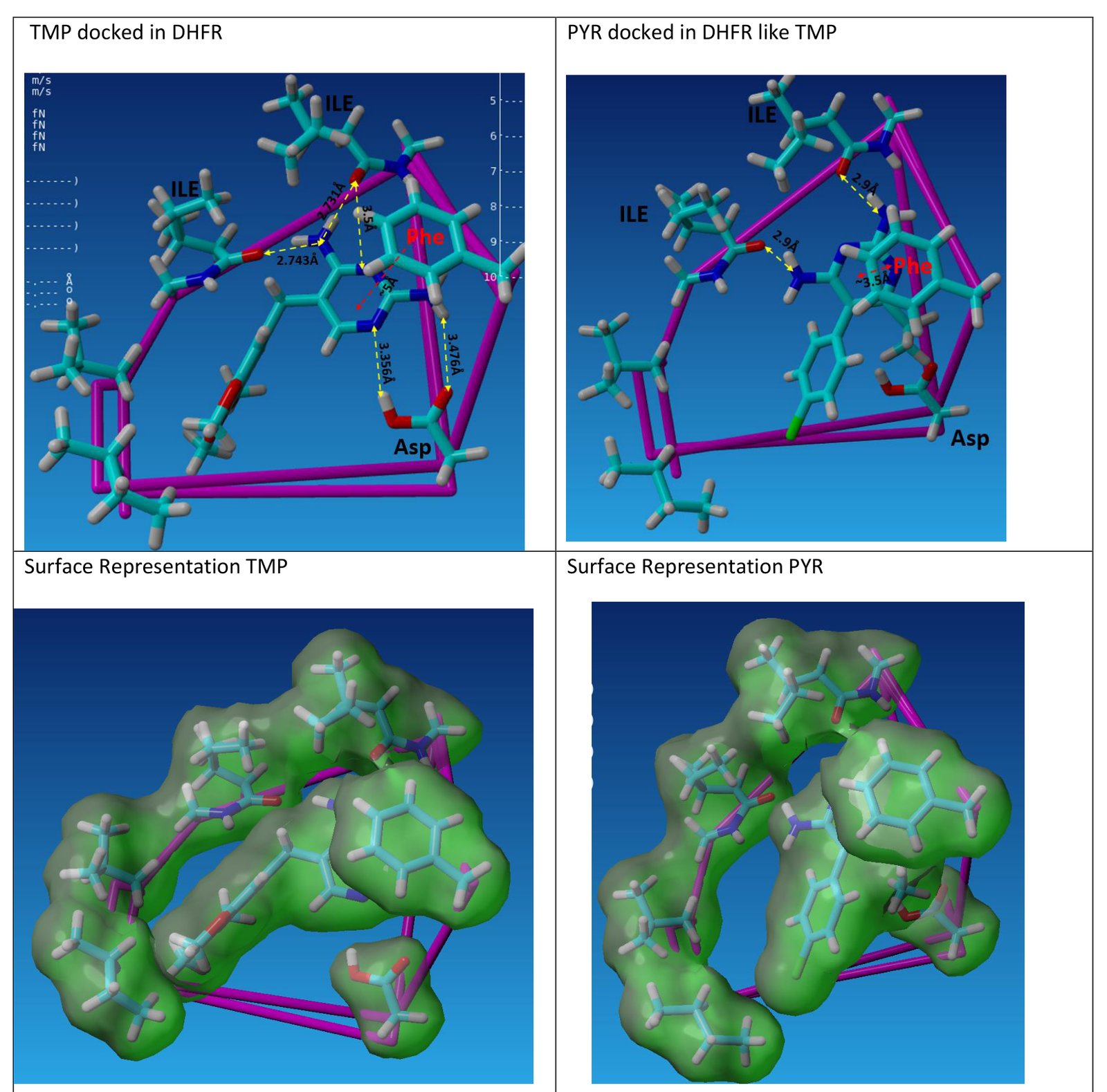
Top: both drugs docked the same way with measured distances. Bottom: surface views, where pyrimethamine’s overlap with the site shows directly why it cannot bind as trimethoprim does.
Methotrexate makes up to eight hydrogen bonds, six from the pteridine ring and two from the glutamate tail, against four for trimethoprim.
Binding energetics
From dG = -RT ln Ka at 310 K, methotrexate (Ka 2 x 10^8 per M) gives -47.4 kJ/mol and folate (1 x 10^5 per M) gives -28.6 kJ/mol, a gap of about 19 kJ/mol. Their hydrogen bond counts are similar, so the difference comes from electrostatics, van der Waals contacts and shape, folate being planar. Counting hydrogen bonds is not the same as predicting affinity.
All docking here was manual, moving and rotating the ligand in YASARA, not an automated docking program with scoring functions.